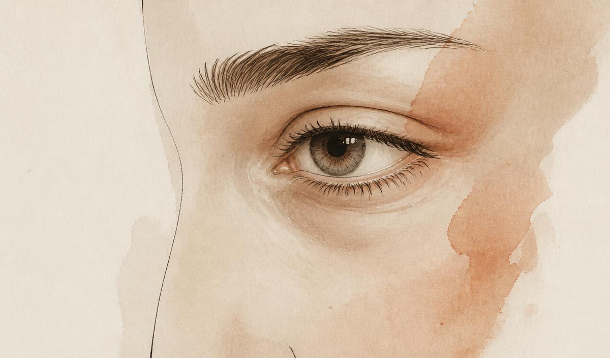
The tear trough is the depression that runs from the inner corner of the eye obliquely along the orbital rim. It is not a wrinkle and not a hollow of "tiredness" — it is a true anatomical transition zone where thin lower-lid skin meets thicker cheek skin over the fixed orbicularis retaining ligament. Every technique that works, works because it respects that anatomy. Every technique that fails, fails because it ignores it.
What actually causes a tear trough
Three structures do almost all the work: the orbital fat that bulges above, the tear-trough and orbicularis retaining ligaments that tether the skin to the maxilla, and the descended malar fat pad that hollows the cheek below. In detail:
- Ligamentous tethering — the tear-trough and orbicularis retaining ligaments pull the skin down to the maxilla.
- Orbital fat pseudo-herniation — the septum weakens and the medial and central fat pads bulge above the trough.
- Midface descent — the malar fat pad slides downward and inward, deepening the trough below.
- Skin quality and pigment — thin, translucent skin transmits the underlying muscle colour, creating the "dark circle" halo.
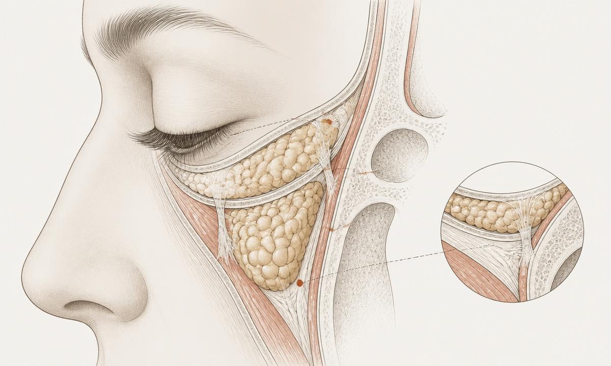
This is why a filler alone almost never gives a clean result in a true ligamentous trough — it inflates the valley without releasing the tether or supporting the cheek above it, which is why long-term results tend to disappoint.
Current treatment strategies
1. Transconjunctival fat repositioning (arcus release)
The current gold standard. A hidden internal incision releases the arcus marginalis and drapes the herniated orbital fat over the orbital rim to fill the trough with the patient's own tissue. No external scar, no fat removed unnecessarily, and the correction is structural rather than volumetric.
2. Extended SOOF lift and midface repositioning
For patients with true midface descent, repositioning the sub-orbicularis oculi fat (SOOF) upward re-establishes the convex lid–cheek junction. It is often combined with fat repositioning above and canthal support laterally.
3. Micro-fat and nano-fat grafting to the deep medial cheek
Small-parcel autologous fat placed deep to the orbicularis — never superficial in the trough itself — restores midface volume and improves skin quality via the regenerative fraction. Nano-fat is used specifically for the pigmented, thin lid skin.
4. Lateral canthal support
Any lower-lid work in a patient with even mild laxity must be paired with a canthopexy or canthoplasty. Skipping this step is the single most common cause of post-operative scleral show and rounding of the eye.
5. Adjunctive skin resurfacing
Fractional CO2 or erbium resurfacing of the lid skin at the same session addresses the pigment and crepe component that no repositioning can fix on its own.
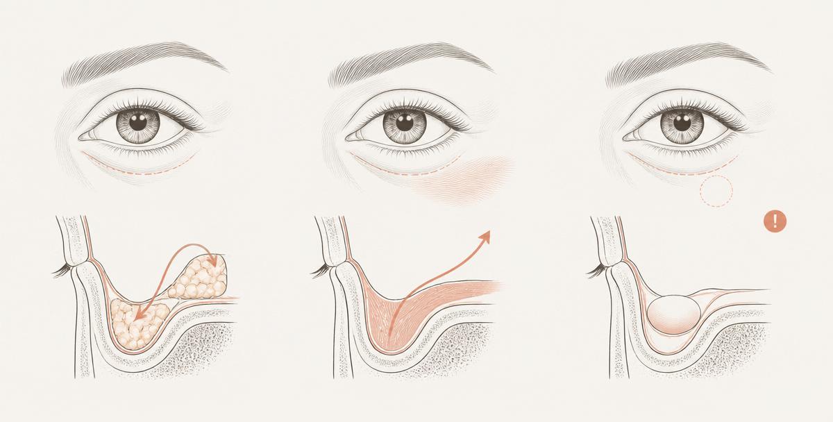
Techniques that are being retired
- Aggressive transcutaneous fat excision — creates the hollow, skeletonised eye that ages badly.
- Superficial hyaluronic-acid filler in the trough — Tyndall effect, chronic swelling, and migration reported years later.
- Isolated lower blepharoplasty without canthal support — high revision rate for scleral show and ectropion.
Choosing the right technique — a quick matrix
| Primary finding | Preferred approach |
|---|---|
| Fat herniation with a mild trough, good skin | Transconjunctival fat repositioning |
| Deep ligamentous trough, minimal fat | Ligament release with deep-plane fat graft |
| Midface descent with cheek hollowing | SOOF lift with fat repositioning and canthopexy |
| Excess skin, crepe, festoons | Skin-muscle flap blepharoplasty with fractional resurfacing |
| Mild, volume-only trough with good skin | Conservative filler, deep and supraperiosteal |
| Pigment-dominant "dark circles" only | Nano-fat and topical protocol, no surgery |
"The tear trough is not corrected by adding volume. It is corrected by releasing what tethers the skin and repositioning what has descended. Structural problems need structural solutions — anything less is cosmetic camouflage."
This article is for educational and informational purposes only and does not constitute medical advice, diagnosis, or treatment. Individual results vary and all surgical procedures carry risks, which are reviewed during an in-person consultation with a qualified physician.
Considering this procedure?
Book a private consultation with Prof. Dr. Mehmet Veli Karaaltın for a personalised surgical plan.
Speak to the clinic
Patients travel from the UK, Ireland and the United States for treatment in Istanbul. Video consultations are held before you book any travel.
Teşvikiye, Sakayık Sk. No:47
34365 Şişli / Istanbul, Turkey